Ethics statements
The experimental procedures involving mice had been performed in strict compliance with the laws governing animal welfare ethics established by the Experimental Animal Middle of Kunming College of Science and Expertise. These procedures had been totally reviewed and accepted by the Animal Ethics Committee (Permitted No. PZWH (Yunnan) K2022-0019). All cell experiments involving DMD sufferers had been accepted by the Medical Ethics Committee of the Third Medical Middle of the Chinese language PLA Common Hospital (Permitted No. KY2121-001). Knowledgeable consent types, accepted by the Ethics Committee, had been signed by the mother and father or guardians of underage individuals. All procedures involving NHPs had been performed in strict compliance with the moral necessities for experimental animals outlined by the Laboratory Animal Ethics Committee of Kunming College of Science and Expertise (Approval Quantity: KUST202301010).
Mice
mdx (C57BL/10ScSn-Dmdmdx/J) mice had been generously offered by Professor Yafeng Music’s laboratory on the Institute of Sport and Well being Science, Beijing Sport College. DmdΔEx50 (C57BL/10-Dmd
NHPs
DMD is an X-linked recessive genetic dysfunction that primarily impacts males. On this examine, 4 wholesome 1.5-year-old male cynomolgus monkeys had been chosen because the analysis topics. These animals confirmed no indicators of an infection or different hostile circumstances. The titers of NAbs in opposition to MyoAAV of their serum had been all beneath 1:5. The monkeys had been randomly assigned to 2 teams: the MyoAAV injection group and the saline management group. All monkeys used within the experiments had been housed within the animal facility of the Institute of Primate Translational Drugs at Kunming College of Science and Expertise. The room temperature was maintained at 22 ± 1 °C with roughly 50% humidity. The indoor lighting schedule was mounted from 8:00 a.m. to eight:00 p.m. The monkeys had advert libitum entry to water and had been offered three meals every day, consisting of fruits and a balanced compound feed.
Cell strains, cell tradition, and transfection
The HEK293T (SCSP-502) and Neuro2a cell strains (SCSP-5035) had been obtained from the Nationwide Assortment of Authenticated Cell Cultures and cultured in DMEM medium supplemented with 10% fetal bovine serum (FBS) and 1% penicillin-streptomycin (PS) at 37 °C in a humidified incubator with 5% CO2. Plasmid transfection was carried out utilizing Lipofectamine 3000 (Thermo Fisher, # L3000015), and cells had been collected for RNA extraction 48 hours post-transfection. Skeletal muscle cells from cynomolgus monkeys had been remoted and cultured following the protocol described in Strategies in Molecular Biology51.
PBMCs from two male DMD sufferers had been remoted from the peripheral blood utilizing 15 ml SepMate tubes (STEMCELL, #15450) and Lymphoprep density gradient medium (STEMCELL, #07801). All cell culture-related experiments involving DMD affected person’s PBMCs had been performed in compliance with the Chinese language PLA Common Hospital Ethics Committee tips (Approval quantity KY2121-001). iPSCs had been generated from PBMCs utilizing the Invitrogen Sendai virus package (Invitrogen, #A16518). The wholesome management group cell line H9 (SCSP-307) had been obtained from the Nationwide Assortment of Authenticated Cell Cultures in China. Differentiation of iPSCs into skeletal muscle was carried out following the flowchart outlined in Fig. 5c. The medium used at completely different tradition phases are listed in Supplementary Desk S1. For myotube formation, myoblasts had been grown in 12-well plates to 90% confluence earlier than switching to myotube differentiation medium. Two days post-differentiation, MyoAAV-UA (2 × 10¹¹ viral particles) was added to every effectively. The medium was changed 24 hours after transduction, and cells had been harvested 5 days post-infection for mobile immunostaining, RNA and protein extraction, and subsequent evaluation.
Teratoma assay
iPSC clones had been indifferent by digesting with 50 μl of 20 mg/ml sort IV collagenase in 1 ml of medium at 37 °C for 10 minutes. The tradition dish was gently tapped to launch the clones, and the supernatant was transferred to a 15 ml tube. After settling for 20 seconds, the supernatant was discarded, and the cells had been washed twice with DPBS. The clones had been additional digested with 1 ml of Accutase (STEMCELL, #07920) at 37 °C for 10 minutes, neutralized with 1 ml of medium, centrifuged at 300 × g for five minutes, and resuspended. After counting, 1 × 10⁶ cells had been blended with 100 μl of Matrigel (Corning, #354277) in a 1.5 ml tube. The cell suspension was injected subcutaneously into NOD-SCID IL2Rγnull mice, guaranteeing no blood within the syringe. Tumor formation was monitored, and when the teratoma dimension reached 3, the tumor was excised, mounted, and stained to guage the pluripotency of the iPSCs.
Plasmid development
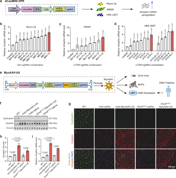
The sgRNAs had been designed primarily based on the PAM web site sequence TTTR utilizing the CRISPR RGEN Instruments (www.rgenome.web), as detailed in Supplementary Information 1. To assemble the dCasMINI-VPR-U6-crRNA scaffold vector, the dCasMINI-VPR sequence was obtained from the plasmid pHR-PGK-SV40_NLS-dCasMINI-V4-VPR-c-Myc_NLS-mCherry-WPRE (Addgene: pSLQ9926), and the DNA fragment was subcloned into an AAV vector. The dCasMINI and VPR components had been underneath the management of a CMV promoter, and the human U6 promoter was related to the activation factor to manage the sgRNA and its crRNA. ClonExpress MultiS One Step Cloning Equipment (Vazyme, #c113-01) was used for ligation. For the development of the vector containing a muscle-specific promoter and twin sgRNAs, pX601-AAV-SPc5-12-dCasMINI-VPR-Cmyc-U6-M8-M12 and pX601-AAV-SPc5-12-dCasMINI-VPR-Cmyc-U6-H1-H10 had been designed as built-in vectors for MyoAAV packaging. These plasmids included a muscle-specific promoter (SPc5-12) and twin sgRNAs focusing on the utrophin gene.
AAV packaging and injection in Dmd mice
MyoAAV was packaged by PackGene Biotech (Guangzhou, China). Two-week-old male mdx mice and DMDΔEx50 mice had been administered the virus both by native injection or tail vein intravenous injection, respectively. Management mice (2-week-old male C57BL/6 mice) had been injected with an equal quantity of saline. A minimal of six mice had been used for every experimental situation.
Immunosuppressant administration and intravenous MyoAAV-UA injection in NHPs
The oral administration of immunosuppressant routine was tailored from the AVXS-101-CL-303 scientific trial (ClinicalTrials.gov identifier: NCT03306277), a Part 3 examine evaluating the efficacy and security of Onasemnogene abeparvovec (AVXS-101) in sufferers with Kind 1 SMA. Within the current examine, prednisolone was administered orally at 1 mg/kg/day beginning 5 days earlier than the injection and continued for 30 days. The dose was then tapered to 0.5 mg/kg/day for 2 weeks, adopted by 0.25 mg/kg/day for a further two weeks. Two wholesome 1.5-year-old male monkeys obtained a systemic dose of MyoAAV-UA at 5 × 10¹³ vg/kg, packaged by PackGene Biotech (Guangzhou, China). The injection was administered by the saphenous vein in the precise decrease limb, with the virus diluted in 30 ml of saline and infused over 30 minutes, adopted by a ten ml saline flush. The management group consisted of two wholesome age-matched male monkeys, which obtained the identical quantity of saline infused on the similar fee because the MyoAAV-UA group. Put up-injection, all monkeys had been monitored for no less than 30 minutes, with no irregular indicators noticed throughout this era.
AAV genome copy numbers and vector genome quantification in tissue samples
Genomic DNA was extracted from tissues utilizing the Wizard® Genomic DNA Purification Equipment. DNA focus and purity had been measured utilizing a Nanodrop 2000 spectrophotometer, and samples had been diluted to 100 ng/µl. AAV viral copy quantity in tissues had been quantified utilizing qPCR with SYBR Inexperienced Expertise and QX200™ AutoDG™ Droplet Digital™ PCR. A 22 µL response combination was ready with QX200 ddPCR EvaGreen Supermix and 100 nM primers. DNA samples had been diluted to 50 ng/µl and pre-treated with DNaseI. The 22 µL response combination was transferred to a 96-well plate, sealed, and subjected to PCR. Outcomes had been routinely analyzed utilizing Quanta software program. The Variety of vector genomes (dCasMINI) per diploid genome (Gapdh) was decided utilizing qPCR (Supplementary Desk S2). Absolute quantification of dCasMINI and GAPDH/Gapdh in every pattern was achieved utilizing normal curves generated by amplifying plasmids containing dCasMINI or GAPDH/Gapdh sequences. These normal curves had been used to calculate the ratio of vector genomes to diploid genomes.
Blood assortment and serum separation
The forearm or hind leg pores and skin of the monkey was shaved and disinfected with iodine. Utilizing a vacuum blood assortment tube (BD, #367983), 3 ml of blood was collected from both the brachial vein or small saphenous vein. The blood assortment tube was left undisturbed for 30 minutes to permit clot formation. Afterward, the tube was centrifuged at 3000 × g for 10 minutes. The pale-yellow serum layer was fastidiously aspirated utilizing a sterile, enzyme-free pipette and saved at −80 °C for long-term use.
ELISA dedication of mouse serum CK
Serum CK in mice was measured utilizing the CK Exercise Assay Equipment (Colorimetric) (Abcam, #ab155901), following the producer’s directions for correct quantification.
MyoAAV NAb assay
The MyoAAV4E-CMV-β-galactosidase virus (1 × 10¹³ vg/ml) was packaged by Guangzhou PackGene Biotech. For every effectively in a 96-well plate, a complete of 109 virus genome copies had been calculated. Combine 10 µl of the virus inventory with 5500 µl of diluent (2% FBS + 97% DMEM + 1% PS). Carry out gradient dilution (e.g., 1:2, 1:4, 1:8) and blend 55 µl of every dilution with 55 µl of virus diluent. Incubate the mixtures at 37 °C for 1 hour. Add the combination to a 96-well plate seeded with HEK293T cells at 60% confluence. Every pattern is examined in duplicate. Embody three optimistic management wells containing the virus diluent solely and three detrimental management wells containing basal medium solely. Incubate the plate at 37 °C with 5% CO2 for 72 hours. After incubation, add 100 µl of X-Gal cell lysis resolution to every effectively, let it sit for 10 minutes, and pipette gently to disperse the cells. Switch 50 µl of the lysate to a 96-well plate, add 50 µl of β-galactosidase assay reagent (Beyotime, # C0602), and incubate at 37 °C for 1 hour. Add 150 µl of response termination resolution to every effectively and measure the absorbance at 420 nm utilizing a plate reader. The NAby titer for every pattern is decided by figuring out the dilution at which the OD worth is half the common of the optimistic management wells. This absorbance corresponds to the pattern’s NAb exercise.
Blood biochemical index testing
After sampling, serum samples are saved in a 4 °C fridge and delivered to the biochemical testing division of Kunming Biotech Worldwide Co., Ltd. on the identical day. Blood biochemical index (ALT, AST, Creatinine, GGT, CHO, Glucose) testing is performed utilizing the Roche C511 automated biochemical analyzer.
Entire blood cell evaluation
Anticoagulated complete blood is collected utilizing EDTA-K2 vacuum blood assortment tubes (BD, #367863). 1 ml of blood collected from the experimental monkeys was transferred to the biochemical testing division of Kunming Biotech Worldwide Co., Ltd. Inside 2 hours, pink blood cells, white blood cells, platelets, and so forth., had been analyzed utilizing the Sysmex XN-1000 automated hematology analyzer from Sysmex Company.
Mice tissue procurement and storage
Below laws governing animal welfare, euthanasia was carried out on the mice to attenuate ache and misery. Subsequently, the center, gastrocnemius (GS) muscle, QDs, diaphragm, liver, kidneys, lungs, mind, and spleen had been collected and additional processed. The muscle groups had been wrapped with yellow beeswax and flash-frozen in liquid nitrogen-precooled isopentane for 30 seconds to keep up muscle cell morphology. They had been then instantly transferred to dry ice and transported to a −80 °C freezer for storage. The remaining tissues had been embedded in OCT and positioned on dry ice for pre-cooling embedding. All samples had been thawed for no less than 1 hour in a −20 °C freezer earlier than sectioning to protect tissue morphology. The sections had been promptly saved in a light-protected −20 °C freezer. Samples for Western blot evaluation and RNA evaluation had been collected in 1.5 ml RNase-free EP tubes, sealed tightly, and flash-frozen in liquid nitrogen for preservation. They had been then transferred to a −80 °C freezer for long-term storage.
RNA evaluation and Sanger sequencing
The full RNA of cells and tissues was extracted utilizing TRIzol (Invitrogen, # 15596026), and 1 μg of RNA was reverse-transcribed to cDNA with the PrimeScript™ (Takara, # RR047) RT package. The TransStart® Inexperienced qPCR SuperMix Equipment (Transgen, # AQ101-01) is used to quantify the relative mRNA expression of genes. Sanger sequencing was carried out by Tsingke Biotechnology Co., Ltd. The primers are proven in Supplementary Desk S2.
Transcriptome evaluation
The mouse GS muscle was shortly frozen with liquid nitrogen, and the tissue block was floor in liquid nitrogen to stop protein and RNA from being degraded. The full tissue RNA was extracted with TRIzol and the mRNA was enriched by oligonucleotide (dT) magnetic beads to assemble a cDNA library. Nanodrop 2000 was used to find out the focus of nucleic acid. Illumina sequencing platform was used for high-throughput sequencing, and the sequencing learn size was PE150. Customized R scripts had been used to carry out additional TPM (transcripts per million mapped reads) normalization and high quality management. Downstream plots used the ggplot252. Sequencing knowledge had been matched to Mus musculus genome knowledge (GRCm39) utilizing hisAT2 (Model 2.2.1). Transcriptome meeting utilizing a hybrid of lengthy and quick reads with StringTie53. Moderated estimation of fold change and dispersion for RNA-seq knowledge with DESeq2 (biomedcentral.com). GO pathway enrichment evaluation elucidates the connection between utrophin gene expression and the regulation of related pathways54. The WGCNA package deal supplies R capabilities for weighted correlation community evaluation for use for locating clusters (modules) of extremely correlated genes55. The Random Forest package deal supplies a Rinter-face to the Fortran applications by Breiman and Cutler (obtainable at http://www.stat.berkeley.edu). To carry out the Gene Set Variation Evaluation, the GSEABase package deal (model 1.44.0) was used to load the gene set file, which was downloaded and processed from KEGG database (https://www.kegg.jp/). To assign pathway exercise estimates to particular person cells, we utilized GSEA utilizing normal settings, as applied within the GSEA package deal (model 1.30.0). The variations in pathway actions scored per cell had been calculated with the LIMMA package deal (model 3.38.3)56.
Western blotting
Myocytes had been digested with 0.05% trypsin and picked up by centrifugation at 300 × g. Mouse or NHP muscle tissue was frozen in liquid nitrogen and floor into powder. RIPA lysis buffer with PMSF was added to extract protein from the cell pellet or tissue. The combination was lysed on ice for 40 minutes, vertexing each 5 minutes. Protein supernatant was obtained by centrifuging at 10,000 × g for 10 minutes at 4 °C. Proteins had been separated by SDS-PAGE and transferred to Immobilon-P® membranes. Membranes had been blocked with 5% skim milk, incubated with antibodies in a single day, washed, and detected utilizing ECL substrate. Band depth was analyzed with ImageJ. The first antibodies are proven within the reporting abstract.
Histological evaluation
All muscle groups embedded in yellow beeswax had been cryo-sectioned at 8 µm. Hematoxylin-eosin staining (Solarbio #G1120) and Sirius pink staining (Solarbio, #G1472) had been performed in keeping with the producer’s directions. DAB (3,3’-Diaminobenzidine) staining for muscle sections was performed following the protocol outlined within the Beyotime Peroxidase (BSP) Immunohistochemistry Equipment (Beyotime #P0202).
Immunofluorescence staining
Cultured cells, skeletal muscle groups (QDs, brachialis, and diaphragm), and hearts had been cryo-sectioned at 8 µm and stuck with 4% paraformaldehyde for 10 min. Sections had been blocked and permeabilized with 3% BSA and 10% FBS containing 0.2% TritonX-100 for two hours at room temperature. Sections and dishes had been then incubated with major antibody in a single day at 4 °C, washed with 1× PBST 3 times, 5 minutes every, adopted by donkey anti-rabbit/mouse IgG H&L secondary antibody for two hours at room temperature. Use TureBlack (Biotium #23007) to scale back autofluorescence in tissue sections in keeping with the post-treatment with TrueBlack following immunostaining. Slides had been mounted with an antifade mounting medium containing DAPI (Abcam, #ab104139). Immunofluorescence pictures had been taken with Nikon AX and Leica SP8 confocal microscope. The first antibodies are proven within the reporting abstract.
Morphometric evaluation
Three teams of mice, C57BL/6 (wildtype) and mdx randomized to both injection with saline or MyoAAV-UA, had been studied by investigators blinded to specimen identification (n = 6 mice for every group). Three teams of mice brachialis, and QDs had been stained with H&E screened with a lightweight microscope. six muscle sections from completely different mice per group had been evaluated for the centrally nucleated fibers. Areas on the myotendinous junctions had been excluded from the measurements as they’re wealthy in each mdx and controls. In complete, 7439 fibers had been evaluated for centrally nucleated fibers share. And the share of fibrotic space in brachialis, QDs, diaphragm, and coronary heart sections staining with Sirius Crimson Staining. six muscle sections from completely different mice per group had been evaluated for fibrotic space share in ImageJ software program, every muscle part was measured in 3 mm2 per muscle. The realm of curiosity (AOI) instrument is employed to manually define all full muscle fibers. Within the HSI mode, utilizing the H/S/I curve instrument, the muscle fibers are labeled as pink, whereas the interstitium is left unlabeled. Subsequently, the full space of muscle fibers inside the AOI is measured. Six sections of the brachialis and QDs muscle groups are randomly chosen for statistical evaluation in every group of mice.
Behavioral evaluation
Mouse Gripper (Shanghai Mei Rui Sai Scientific Instrument Co., Ltd. #MC-RMG01) was used to measure the mice forelimb greedy drive. The tester gently pulls the mouse’s tail along with his hand to make the mouse’s hind limbs hold within the air whereas the forelimbs grasp the barbed wire to check its energy. The physique of the mouse is parallel to the barbed wire. Every measurement was carried out 10 occasions, and the common worth was used for statistical evaluation. The Rotarod-Lantency to fall experiment is used to guage the train endurance of mice (Shanghai Mei Rui Sai Scientific Instrument Co., Ltd. #MC-RMM02). Referring to the experimental design scheme of the Rotor-Rod of the Behavioral and Useful Neuroscience Laboratory of Stanford College, the mice had been subjected to a one-week coaching, to make sure that all topics discovered the duty to the identical diploma. Mice in every group had been examined on the similar pace because the working wheel, and the time for the mice to fall from the wheel was recorded, every mouse ran three rounds for statistical evaluation. Every mouse was positioned within the Open Subject equipment (40 × 40 cm) and recorded for 30 minutes at evening. All mice might be pre-exposed to the open area setting for 10 minutes. The motion and distance of mice had been analyzed by Good v3.0 software program.
IFN-γ assay
Following the directions outlined within the Thermo Fisher Rhesus Macaque IFN-gamma ELISA Equipment (Thermo, # EP8RB) protocol, we performed the detection of IFN-γ ranges in serum samples. After equilibrating the serum to room temperature, we diluted it twofold with 1× Assay Diluent earlier than continuing with the detection. The IFN-γ values within the serum had been then calculated utilizing the usual curve.
Ultrasound examination
Animals had been administered 0.1 mg/kg of atropine and 0.05 ml/kg of Zoletil intramuscularly for anesthesia. The procedures had been swiftly carried out after inducing anesthesia. A coupling agent was utilized to the examination web site to get rid of air obstruction between the probe and the pores and skin, lowering friction between them. Appropriate probes of the LOGIQ Fortis Professional ultrasound machine had been chosen for analyzing the carotid arteries, thyroid gland, liver, kidneys, coronary heart, and different areas. Following the examination, animals had been administered native muscle injection of atipamezole 0.05 ml/kg for reversal of anesthesia. Animal circumstances had been noticed for no less than 30 minutes, feeding and watering had been resumed 4 hours after awakening from anesthesia.
Mind MRI
Animals had been administered 0.1 mg/kg of atropine and 0.05 ml/kg of Zoletil intramuscularly for anesthesia. They had been positioned in the precise lateral decubitus place for the process. Utilizing a SIEMENS Prisma 3.0 T MRI scanner, the top coil was chosen for scanning. Imaging knowledge included T1-weighted pictures and T2-weighted pictures. The essential parameters for knowledge acquisition had been as follows: 3D T1 sequence: TR/TE = 1400 ms/3.55 ms, inversion time = 900 ms, flip angle = 8 levels, area of view = 135 × 135 mm2, matrix dimension = 224 × 224, slice thickness = 0.6 mm, voxel dimension = 0.6 × 0.6 × 0.6 mm3, sections = 160; acceleration issue = 2; NEX = 3. 3D T2 sequence: TR/TE = 2500 ms/520 ms, area of view = 115 × 115 mm2, matrix dimension = 192 × 192, slice thickness = 0.6 mm, voxel dimension = 0.6 × 0.6 × 0.6 mm3, sections = 128; acceleration issue = 2; NEX = 4.
Muscle sampling and evaluation of NHPs
In a sterile working room, cynomolgus monkeys had been anesthetized with intramuscular injections of 0.1 mg/kg atropine and 0.05 ml/kg Zoletil. Injections had been administered away from the surgical web site. The pores and skin of the legs or arms was shaved and disinfected with iodine. A sterile drape uncovered the surgical space. Utilizing sterilized devices, the pores and skin and fascia had been incised, avoiding main blood vessels. Hemostatic forceps separated the muscle fascia, and the muscle was lower to acquire tissue for evaluation. Hemostasis was achieved with electrocautery or ligature, and the layers had been sutured. The realm was disinfected and dressed. Postoperative awakening remedy, atipamezole (0.05 ml/kg) was administered intramuscularly. Previous to AAV therapy, biopsies had been collected from the precise QDs of 4 cynomolgus monkeys. 4 weeks post-treatment, biopsies had been collected from each the left GS and left BB muscle groups of the MyoAAV-UA handled and saline management monkeys. The monkey’s restoration was monitored for no less than 30 minutes, and feeding and watering had been resumed 4 hours after awakening from anesthesia. Ceftriaxone (50 mg/kg/day) was administered for 3 days to stop an infection.
Bone density evaluation (iDXA) and chest X-ray scan evaluation
Following the anesthesia depicted earlier than, animals had been positioned in a supine place for X-ray examination of the chest utilizing the X-ray HPS-HF-4.0 system (SEDECAL, SPAIN) to research lung texture distribution and cardiac dimension. Entire-body scans had been carried out utilizing the Lunar iDXA system (GE Healthcare) to measure complete physique fats, lean muscle mass, and fats distribution ratio. The evaluation was performed utilizing the next software program variations: DPXL 4.7e, Prodigy 8.80, and iDXA 10.40. Particular anatomical landmarks described beforehand had been used to partition areas, together with the arms, legs, and trunk. For high quality management of soppy tissue mass associated to densitometry, scans had been carried out month-to-month utilizing 8-liter bottles crammed with methanol and water to simulate fats and non-fat comfortable tissue, respectively. iDXA and X-ray assessments had been performed in Kunming Biotech Worldwide Co., Ltd. MRIcroN model 4.0 software program is used to research mind MRI pictures.
Temperature assay
Utilizing the infrared digital thermometer (CEM, #DT-8806H), align it with the chest pores and skin of the cynomolgus monkey, guaranteeing that the gap between the detector and the pores and skin doesn’t exceed 2 centimeters. Take three consecutive measurements, document the values, and calculate the common.
Statistical evaluation
For the mouse experiments, outcomes had been derived from n = 6 organic replicates. For the NHP experiments, n = 2 organic replicates had been used. Myoblasts had been differentiated from two impartial iPSCs organic samples with completely different DMD gene mutation sorts and the H9 cell line. Statistical evaluation was carried out for knowledge with a organic pattern dimension higher than 3. One-way ANOVA adopted by the Tukey submit hoc check was used for knowledge with three teams. Statistical graphs had been plotted by GraphPad Prism (model 8.0.2) software program. Pattern sizes and p values are offered within the Determine legends and Supply Information file. P values smaller than 0.05 had been thought of statistically important. Western blotting quantification, centrally nucleated myofibers, and Sirius red-stained muscle fibrosis had been analyzed utilizing ImageJ software program (model 1.53k). Sanger sequencing outcomes had been aligned utilizing SnapGene software program (model 4.2.4). Immunofluorescence staining pictures had been analyzed utilizing NIS factor viewer (model 5.21) and Leica Software suite X (model 3.7.4). Histopathological staining pictures had been analyzed with Olyvia software program (model v3.2). Droplets digital PCR (ddPCR) occasions had been analyzed utilizing Quanta software program (model 1.7.4). Absorbance measurements had been carried out utilizing SpectraMax software program (model 7.1). X-ray examination of the chest was performed utilizing the X-ray HPS-HF-4.0 system (SEDECAL, SPAIN). Bone density evaluation had been carried out utilizing DPXL (model 4.7e), Prodigy (model 8.80), and iDXA (model 10.40). Ultrasound was carried out suing The LOGIQ Fortis Professional Ultrasound System.
Reporting abstract
Additional data on analysis design is on the market within the Nature Portfolio Reporting Abstract linked to this text.