Molecular cloning
The DNA sequence harboring the 6x Histidine tag, the T7 peptide chief, a TEV proteolytic cleavage web site, and eGFP (6xHis-T7Leader-TEV-eGFP) was remoted from Addgene assemble (#54762). eGFP was cloned right into a pET21a(+) vector (EMD Millipore #69740) spine by restriction enzyme digest adopted by T4 DNA ligation. DNA sequence was confirmed through Sanger Sequencing (Eurofins).
The DNA sequence harboring the 6x Histidine tag, the T7 peptide chief, an X-press affinity tag, and mStayGold (6xHis-T7Leader-XpressTag-mStayGold) was amplified from Addgene assemble (#212017). The DNA sequence harboring the 6x Histidine tag, the T7 peptide chief, a TEV proteolytic cleavage web site, and mTagBFP2 was amplified from Addgene (#54572). The fragments have been assembled through Gibson meeting into the pRSETB vector spine (Addgene #212017)28,29. DNA sequences have been confirmed through Sanger Sequencing (Eurofins).
BB recipe
The BB recipe used for all experiments on this examine consists of the next:
12 g/L tryptone (Millipore Sigma T9410)
24 g/L yeast extract (Millipore Sigma Y1625)
KH2PO4 (0.05–0.1 M)
Na2HPO4 (0.05–0.1 M)
NH4Cl (0.05 M)
Na2SO4 (0.005 M)
MgSO4 (0.002 M)
Glycerol (0.54 M)
D-glucose (0.03 M)
D-galactose (0.055 M)
Na2C6H6O7 (0.025 M)
FeCl2·6H2O (0.1 M)
CaCl2 (0.02 M)
MnCl2·4H2O (0.035 M)
ZnSO4·7H2O (0.087 M)
CoCl2·6H2O (0.0087 M)
CuCl2·2H2O (0.002 M)
NiCl2·6H2O (0.002 M)
Na2MoO4·5H2O (0.002 M)
FeSO4·7 H2O (0.0179 M)
(NH4)6Mo7O24·4 H2O (0.0009 M)
H3BO3 (0.186 M)
EDTA (0.05 M)
CuSO4·5 H2O (0.0062 M).
The pH of the media is adjusted to the impartial vary (7.3–7.6).
eGFP induction and in-cell eGFP measurements
In-cell eGFP fluorescence depth measurements have been carried out as beforehand described with minor modifications14. eGFP expression assemble was reworked into BL21(DE3) competent cells (New England Biolabs). An in a single day starter tradition was grown in LB at 37 °C with 100 ug/mL carbenicillin. 50 mL manufacturing cultures in non-baffled disposable cell tradition Fernbach flasks (Corning 431143) have been began in every media (LB, TB, MM, ZYM, BB) with 100 ug/mL carbenicillin by 1:100 dilution of the starter tradition and have been grown at 30 °C at 250 rpm. 1 mM IPTG was added to LB and TB media at 6 h. For every organic replicate, and every respective time level throughout all 5 examined media, 1 mL of tradition was collected and pelleted at 10,000g for 10 min. Media was decanted, and pellets have been resuspended with 1 mL of 137 mM NaCl, 2.7 mM KCl, 10 mM Na2HPO4, and 1.8 mM KH2PO4 (1x PBS) every. Samples have been diluted with 1x PBS into vary for correct OD600 measurements. Absorbance at OD600 and eGFP fluorescence depth (Ex485/Em516) have been then measured by including 350 µL of diluted pattern right into a 96-well plate (Grenier 655180) and studying in a Synergy H1 plate reader (Agilent). Clean measurements have been subtracted previous to correcting for dilution. 20 mL from all organic replicates, for every kind of media have been individually spun down at 4,000 rpm on the final time level, 36 hours (Fig. 1a). Media was decanted, and tubes have been saved at − 80 °C till eGFP purification and solubility experiments have been executed.
eGFP purification and yield quantification
Cells from a single replicate every of the 5 E. coli eGFP liquid cultures (BB, LB, TB, MM and ZYM) have been resuspended in lysis buffer: 1 mg mL− 1 lysozyme (Thermo Fisher) and 1 mg mL− 1 DNase I (Thermo Fisher) in 1x PBS containing 5 mM imidazole. Cells have been incubated for 20 min on a nutator, spun down at 17,000 g, 4 °C for 10 min, and clarified protein-containing lysate was collected. 5 cycles have been then carried out wherein the remaining cell pellet was refrozen at − 80 °C, thawed, resuspended in lysis buffer, spun down and the soluble fraction collected. All soluble fractions have been pooled. The pooled lysates from every liquid tradition have been utilized to particular person gravity columns containing 10 mL Ni-NTA resin (Cytiva) equilibrated in 1x PBS. Clarified cell lysates have been flown over every respective gravity column 5 complete occasions at 3 mL min− 1. Gravity columns have been then washed with 1x PBS for > 50 column volumes at 4 °C. Protein elution throughout wash steps was monitored utilizing Bradford reagent (Bio-Rad) till no detectable protein was eluted. eGFP was eluted off Ni-NTA resin with 1x PBS containing 300 mM imidazole pH 7.4. Protein elution from the Ni-NTA resin was monitored by Bradford reagent (Bio-Rad) all through elution. eGFP samples have been buffer exchanged into 1x PBS and concentrated by centrifuge ultrafiltration (Amicon Extremely-15, 10k MWCO, Millipore) to a last quantity of 10 mL every. An aliquot from every pattern was blended 1:1 with Laemmli Pattern Buffer (Bio-Rad), boiled for five min, and loaded right into a 4–20% Mini-PROTEAN® TGX Stain-Free™ Gel (Bio-Rad). Stain-free chemistry incorporates a trihalo compound that covalently binds to tryptophan residues and permits the visualization of proteins following electrophoresis by making the proteins fluorescent when uncovered to UV mild48. The remainder of the samples have been flash frozen with liquid nitrogen for additional evaluation. The stain-free gel was imaged on a BioRadChemiDoc™ MP imaging system and analyzed utilizing ImageLab (v 5.1) software program. Complete intensities for all bands in every lane have been quantified and the fraction of complete protein similar to the eGFP band was decided. Freshly thawed remaining samples have been utilized in a BCA assay49 (Thermo Fisher) to measure the overall protein focus utilizing a Synergy H1 plate reader (Agilent) to measure A562 of 350 µL samples in wells of a transparent 96-well plate (Greiner 655180). The share of eGFP sign measured above was utilized to the overall protein yields measured by the BCA assay. Ultimate corrected eGFP yields have been scaled up from mg/20 mL to mg L− 1 for all 5 media.
Immunoblotting
To detect 6xHis-eGFP within the soluble and insoluble fractions, bacterial lysates both from the soluble fraction of clarified lysate, or from resolubilized insoluble fractions from the cell pellets (8 M urea with buffer trade again to 1x PBS) have been separated on a 4–20% SDS-PAGE gel (Bio-Rad), and proteins have been transferred to Immobilon-P PVDF membrane (EMD Millipore). After blocking in TBS/0.1% Tween 20/5% non-fat dry milk, the blots have been probed with mouse anti-6xHistidine main antibody (Sigma) adopted by the secondary goat anti-mouse IgG2a labeled with HRP (Southern Biotech) diluted in blocking resolution. Blots have been developed utilizing Luminata Crescendo Western HRP substrate (EMD Millipore) and recorded on ChemiDoc MP Imaging System (Bio-Rad).
Human cytochrome c expression and purification
Human cytochrome c (Hu-Cytc) was expressed and purified as described beforehand19. The pBTR (Human Cytc) vector, a spinoff of the pBTR1 expression vector, with the yeast iso-1-Cytc gene changed with an artificial Hu-Cytc gene, was used for expression17,50,51. This vector co-expresses yeast heme lyase, which covalently attaches heme to the Cys-Ser-Glu-Cys-His heme recognition sequence of Hu-Cytc within the cytoplasm of E. coli. Competent E. coli BL21(DE3) cells have been reworked with the pBTR(Human Cytc) vector. The colonies have been washed off the choice plate and grown in 1 L of BB for twenty-four to 36 h at 37 °C with 100 ug/mL carbenicillin. Cells have been then pelleted and suspended in a lysis buffer containing 10 mM Tris pH 7.5, 500 mM NaCl, 2 mM PMSF, and a small amount of DNase. Lysis was carried out through sonication with a Qsonica Q700 sonicator. The lysate was cleared by centrifugation after which incubated in a single day whereas stirring with 12% ammonium sulfate to precipitate impurities. Centrifugation separated out the precipitate, and the supernatant was dialyzed in a single day twice at 4 °C, each occasions in 3 L of buffer containing 12.5 mM sodium phosphate, pH 7.2, 1 mM disodium EDTA, and a pair of mM β-mercaptoethanol (BME). The dialyzed protein resolution was loaded onto a CM Sepharose column that had been equilibrated in 50 mM sodium phosphate buffer, pH 7.0, 1 mM EDTA, and a pair of mM BME. The column was washed with 5 column volumes of this identical buffer to take away any further protein impurities, and the ultimate protein was eluted with a linear gradient from 0 to 0.8 M NaCl in the identical buffer. The eluate was concentrated and exchanged right into a low salt buffer, 50 mM sodium phosphate, pH 7.0, by centrifuge ultrafiltration (Amicon Extremely-15, 3k MWCO, Millipore) earlier than being flash frozen and saved at -80 °C. On the day of the experiment, protein was thawed and additional purified with an AKTA-prime plus chromatography system (GE Healthcare Life Sciences) geared up with a HiTrap SP HP 5 mL cation trade column and a gradient from 0 to 0.3 M NaCl in 50 mM sodium phosphate buffer, pH 7.0, over 30 min at a movement fee of 1 mL/min. The purified protein was concentrated through centrifuge ultrafiltration (Amicon Extremely-15, 3k MWCO, Millipore). Protein yields have been calculated from the ultimate purified, concentrated protein by UV–Vis Spectroscopy previous to peroxidase exercise assays52.
Peroxidase exercise measurements
The peroxidase exercise of human cytochrome c (Hu-Cytc) was measured through conversion of guaiacol to tetraguaiacol which strongly absorbs at 470 nm19,27. Hu-Cytc was oxidized with potassium ferricyanide, Okay3[Fe(CN)6], at room temperature for 7 to 10 minutes, after which the Okay3[Fe(CN)6] was separated from the protein utilizing a G25 Sephadex desalting column (Cytiva) equilibrated with 50 mM sodium phosphate buffer at pH 7.0. The buffer resolution was degassed beneath argon for 30 to 45 minutes to inhibit non-enzymatic oxidation of guaiacol by dissolved O2. Options of 4 μM protein, 400 μM guaiacol, and 100 ± 4 mM hydrogen peroxide have been made within the degassed buffer. The H2O2 focus used was chosen in order that it was above the Michaelis fixed, Okaym, of H2O2 for peroxidase exercise which is about 37 mM for human Cytc21,22,23,24,27,53. Guaiacol and H2O2 concentrations have been measured by absorbance utilizing extinction coefficients, e274 = 2150 M− 1cm− 1, and e240 = 41.5 M− 1cm− 1, respectively25,26,27. The protein resolution, degassed buffer, and guaiacol options in numerous concentrations have been blended instantly previous to taking measurements. The protein/guaiacol resolution was then robotically blended in a 1:1 ratio with the 100 mM peroxide resolution utilizing an Utilized Photophysics SX20 stopped-flow spectrometer. Time-dependent modifications have been recorded at 470 nm (A470). Ultimate protein focus was 1 mM, and last H2O2 focus was 50 mM. All knowledge measurements have been taken at 25 ± 0.1°C. 5 traces have been recorded per guaiacol focus. A470 was plotted as a operate of time for every trial, and the best slope of the linear area was used to find out preliminary enzyme velocity, ν. The slopes have been averaged, then multiplied by 4 since oxidation of guaiacol removes 4 electrons, divided by last protein focus and multiplied by 26.6 mM− 1cm− 1, the extinction coefficient for tetraguaiacol (3,3’-dimethoxy-4,4’-biphenoquinone)19,21,53. This offers ν/[Cytc] values as a operate of guaiacol focus. The Michaelis-Menten equation was match to the info utilizing SigmaPlot v. 13 to extract the Michaelis fixed, Okaym, in addition to the catalytic fee fixed, okcat.
Twin expression of fluorescent proteins in E. coli
BL21(DE3) competent cells have been reworked and cultivated with carbenicillin resistance28. Every flask for every replicate was inoculated with 1:100 starter tradition from in a single day cultivation (14 h) for BB, TB, and LB media. 50 mL complete cultures have been used for every replicate and cells have been grown at 37 °C till reaching OD600 0.6. Then, TB and LB cultures have been induced with 0.1 mM IPTG, and all cultures have been shaken at 25 °C at 250 rpm for 5 days. Cells have been harvested at 4000 rpm, media was decanted, and samples have been flash frozen with liquid nitrogen, then saved at − 80 °C till movement cytometry experiments.
Circulation cytometry
Samples have been analyzed with the Attune NxT (Life Applied sciences, Grand Island, NY, USA) movement cytometer. Ahead and aspect scatter properties have been displayed on a logarithmic scale with the ahead scatter threshold set to 1000 for all samples analyzed. mStayGold inexperienced fluorescent protein was measured with the BL1 laser line, excitation at 488 nm with emission detection at 530 nm with a 30 nm bandpass filter. mTagBFP2 was measured by the VL1 laser line excitation at 405 nm with emission detection at 440 nm with a 50 nm bandpass filter. Thawed E. coli cells have been resuspended in 1x PBS supplemented with 1mM MgSO4 and 0.1 mM CaCl2, and additional diluted 1:50 in PBS. 100 µL of the cell suspension was analyzed at a movement fee of 25 µL/minute for every replicate. Knowledge was analyzed utilizing FlowJo V10.10 (BD Life Sciences, Franklin Lakes, NJ, USA). Cells of all replicates have been gated for intact single cells based mostly on the ahead and aspect scatter (Fig. S3) and common gates for ± mStayGold and ± mTagBFP2 have been set based mostly on the unfavourable management cells cultured in BB, however not expressing fluorescent proteins (Fig. S3).
Imaging E. coli
Stay-cell imaging of the bacterial cells was carried out on a Zeiss LSM 880 laser scanning confocal microscope with a Plan-Apochromat 63x/1.4 NA DIC M27 oil immersion goal. A 405 nm laser was used to excite mTagBFP2 with fluorescence detected from 410 to 498 nm. The excitation for mStayGold was a 488 nm laser with fluorescence being detected from 498 to 598 nm. All cultures have been imaged utilizing the identical settings in ZenBlack 2.3. Most depth projections have been generated from z-stacks of E. coli in every respective medium utilizing ZenBlue 3.6.
eGFP expression measurements utilizing M9 agar plates
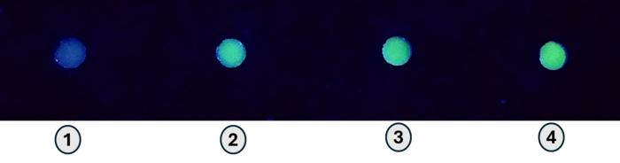
All M9 minimal media mixtures have been ready following the bottom recipe20. Every therapy was supplemented with glucose to a last focus of 0.5% (w/v). The place applicable, further carbon sources (glycerol, glucose, lactose, and galactose) have been additionally added to a last focus of 1% (w/v); for the optimistic management, IPTG was supplemented to a last focus of 0.1 mM. Plates meant for BL21(DE3) moreover contained carbenicillin at a last focus of 100 µg mL− 1, and plates meant for SHuffle® T7 Categorical (New England Biolabs C3029J) contained carbenicillin and streptomycin at last concentrations of 100 µg mL− 1 and 25 µg mL− 1, respectively. Starter cultures of SHuffle® T7 Categorical and BL21(DE3) E. coli have been ready by inoculating eGFP transformants into 30 mL M9 medium (0.5% w/v glucose) containing the suitable antibiotics described above. Flasks have been incubated in rotary shakers set to 250 rpm at 30–37 °C for SHuffle® T7 Categorical or BL21(DE3) E. coli, respectively. The BL21(DE3) tradition was grown for twenty-four h beneath these situations, whereas SHuffle® T7 Categorical wanted 72 h to attain the identical qualitative density. Following this preliminary development, cell suspensions have been plated in parallel on six M9 modifications with the suitable antibiotic choice: M9 (0.5% w/v glucose), M9 + glucose (1% w/v), M9 + glycerol (1% w/v), M9 + lactose (1% w/v), M9 + galactose (1% w/v), and M9 + IPTG (0.1 mM). For every plate, sixteen 10 µL aliquots of homogenous tradition have been dotted in a 4 × 4 grid and allowed to dry beneath a laminar movement hood. All plates have been incubated at 30 °C and have been imaged after 6 days (SHuffle T7 Categorical) or 8 days (BL21(DE3)) of tradition utilizing a BioRad ChemiDoc™ MP imaging system. To evaluate in-colony eGFP fluorescence, photographs of every plate have been captured utilizing a 530/28 filter beneath UV illumination utilizing the identical publicity settings and picture space. Photographs have been analyzed utilizing the companion ImageLab (v5.1) software program, performing automated background subtraction and quantifying built-in colony fluorescence depth for every colony with out normalization for colony measurement. Non-uniform or fused colonies have been excluded from the evaluation, leading to 12–16 values per situation. Common fluorescence depth was calculated for every situation.
Expression and purification of NMDA receptor ABDs
The GluN1 and GluN2A agonist binding domains (ABDs) have been purified utilizing an analogous protocol to beforehand described with a number of changes54. Briefly, the GluN1 S1S2 ABD comprises residues 394–544 and 663–800 from the full-length GluN1 joined by a Gly-Thr dipeptide linker and the GluN2A ABD comprises residues 402–539 and 661–802 from the full-length GluN2A joined by a Gly-Thr dipeptide linker as beforehand described34,54,55. The GluN1 ABD was fused with 6x-Histidine tag and thrombin proteolytic cleavage web site (6xHis-Thrombin-GluN1 ABD)34. The GluN2A ABD consisted of a fusion between 6xHis-small ubiquitin-like modifier and GluN2A ABD (6xHis-SUMO-GluN2A ABD). Every ABD protein was expressed utilizing SHuffle® T7 Categorical E. coli in BB at 25 °C–20 °C for twenty-four h respectively. Every assemble contained a 6xHis-tag on the amino-terminus and was captured by making use of clarified cell lysate to a 5 mL HiTrap IMAC HP column (Cytiva). Following loading of the GluN1 and GluN2A ABDs onto the IMAC, proteins have been eluted with a linear gradient of imidazole concentrations, and peak fractions containing proteins have been pooled and digested with both thrombin protease or ubiquitin ligase protease-1 (ULP-1) to take away N-terminal-tag sequences and isolate GluN1 or GluN2A ABD protein fragments respectively. Put up-cleavage, GluN1/GluN2A ABD proteins have been additional utilized to a 5 mL Hello-Lure SP column (Cytiva) to take away main contaminants, and GluN1/GluN2A ABD protein fragments have been additional purified with Superdex 200 10/300 GL (Cytiva) through size-exclusion chromatography in a buffer consisting of in 10 mM HEPES, 100 mM NaCl, 1 mM glycine, 1 mM L-glutamate, pH 7.0.
Crystallography for GluN1/GluN2A ABDs
Purified GluN1A and GluN2A ABD proteins have been blended at a 1:1 ratio and incubated O/N at 4 °C earlier than the GluN1/N2A advanced was subjected to a different spherical of purification utilizing a Superdex 200 10/300 GL measurement exclusion column (Cytiva) in 10 mM HEPES-NaOH, 100 mM NaCl, 1 mM glycine, 1 mM L-glutamate, pH 7.0. Complexed proteins have been concentrated to 4–7 mg/mL and used to set 1:1 drops in opposition to a reservoir consisting of 0.2 M ammonium acetate, pH 6.8 with 14–18% PEG 4000 utilizing the hanging drop diffusion methodology. Following 48 h of crystal development, two rounds of crystal soaking have been carried out. Within the first spherical of soaking, drops have been soaked with reservoir resolution together with 100 µM glutamate, and 100 µM 7-Chlorokynurenic acid (7-CKA). After at the least 24 h of soaking, the drops have been then soaked for a second time with reservoir resolution together with 100 µM glutamate, and 300 µM 7-CKA for no less than 24 h previous to harvesting. Crystals have been cryoprotected in reservoir resolution containing 20% (v/v) glycerol and flash-frozen in a 100 Okay nitrogen gasoline stream. Diffraction knowledge have been collected on the Stanford Synchrotron Radiation Lightsource (SSRL-SMB) on the 12 − 2 beamline. Photographs have been processed utilizing autoPROC56. Preliminary phases have been decided by molecular alternative in PHASER57 utilizing a printed glycine/glutamate sure GluN1/2A ABD construction (PDB ID 4NF4)34 as search mannequin. The preliminary fashions have been match right into a 2mFo-DFc map with COOT58 and subjected to 1 cycle of inflexible physique refinement utilizing procedures within the PHENIX program suite59. Subsequently, the mannequin was refined by iterative mannequin rebuilding in COOT and refinement with PHENIX. L-glutamate and the 7-CKA ligand have been situated within the construction utilizing the mFo-DFc map60. Knowledge assortment and refinement statistics are proven in Desk S1. Coordinates and diffraction knowledge have been submitted to the Protein Databank with ID 9DA9.
SpCas9 expression and purification
Recombinant nuclease was purified based on a beforehand printed protocol with minor modifications35. Briefly, pET-28b-6xHis-MBP-TEV-Sp-Cas9-siriusGFP-3xNLS (Addgene #78312) was reworked in Rosetta (DE3) pLysS E. coli competent cells (Novagen), and single colonies have been inoculated and grown in a single day. 500mL BB cultures have been inoculated by 1:100 dilution of the starter cultures and grown at 18 °C whereas siriusGFP fluorescence was monitored by time (Fig. S4a). Bacterial pellets have been then re-suspended within the lysis buffer (20 mM Tris [pH 8], 500 mM NaCl, 5 mM MgCl2, and 5 mM imidazole) supplemented with lysozyme (0.5 mg/mL), incubated for 20 min at 4 °C whereas shaking and lysed by high-pressure cell disruption. After clarification by centrifugation (40,000×g, 45 min, 4 °C), the supernatants have been flown over an IMAC 5mL HiTrap 3 occasions in complete for preliminary affinity purification step. Following washes with 50 mL of PBS, SpCas9 was eluted with 400 mM imidazole step gradient, and dialyzed into 20 mM Tris [pH 8], 20 mM NaCl, 5 mM MgCl2 within the presence of TEV protease in a single day at 4 °C. The remainder of the protocol adheres faithfully to35. Based on protocol, two additional steps of purification have been carried out: ion-exchange (IEX) chromatography (HiTrap SP FF column, GE Healthcare) adopted by size-exclusion chromatography (HiLoad 16/600 Superdex 200 PG, GE Healthcare) utilizing an AKTA Pure 25 FPLC system (GE Healthcare). After the ultimate elution, fractions containing SpCas9 have been pooled, concentrated utilizing a centrifugal concentrator (100,000 MWCO) to succeed in at the least 10 mg/mL, and saved in aliquots at − 80 °C till use. The expression and purification was carried out 3 occasions, with the common yield of 8.5 mg/ml.
Preparation of SpCas9-sgRNA RNP for microinjection
For an injection preparation, 11.9 µg Cas9-siriusGFP in 25mM Tris, 300mM NaCl, 2 mM MgCl2, and 1 μm DTT was blended with 2.36 µg sgRNA in TE buffer (1:1 molar ratio), and KCl was added to a last focus of 150mM in a last response quantity of 10 µL. RNP meeting response was incubated whereas being centrifuged (17,000 RCF) for 30 min at 4 °C. For all genome enhancing experiments, ribonucleoprotein complexes (RNPs) have been fashioned between SpCas9 and sgRNA concentrating on coding exon 2 of the ebony (CG3331) locus (Fig. S6a).
Full sgRNA sequence (IDT) with complementary crRNA underlined:
5′CCACAAUUGUCGAUCGUCAGUUUUAGAGCUAGAAAUAGCAAGUUAAAAUAAGGCUAGUCCGUUAUCAACUUGAAAAAGUGGCACCGAGUCGGUGCUUUU-3′.
Drosophila melanogaster microinjection
Wildtype Canton-S flies have been maintained in 6 oz bottles (Flystuff #32-130) with 50 mL commonplace fly meals. Roughly 300 adults aged 2–5 days have been transferred to embryo assortment chambers (Flystuff #59-105) with 2% apple juice agar assortment plates (35 mm petri dish) topped with yeast paste for 30 min. Embryos have been collected, washed with ddH2O, and organized on a 18 × 18 mm coverslip in olive oil. Sutter P-1000 horizontal micropipette puller and Sutter BV-10 Microelectrode Beveler have been used to drag and bevel aluminosilicate glass capillary tubes (Sutter, #AF100-64-10), respectively. Pipettes have been again loaded with injection combine. Syncytial stage embryos have been injected within the posterior finish (Fig. S4C) at 20 °C with a trinocular microscope (ACCU-SCOPE EXI-310) and PM1000 Microinjector (MicroData Instrument Inc.) on handbook setting, facilitated by an N2 gasoline compressor and managed by a hydraulic micromanipulator arrange (Narishige #MMO-4, #MMN-1, #P-12, #GJ-8, #IP). Coverslip with injected embryos was then washed with 95% ethanol and shortly rehydrated with ddH2O. Coverslips have been transferred to meals vials (Flystuff, #32–117) and incubated at 26 ± 2 °C, 65% humidity with a 12 h mild/darkish cycle.
Drosophila crosses and phenotypic characterization
Particular person injected surviving adults have been crossed to three double balanced ebony flies (w[*]; CyO/sp; TM2, es/TM6b, e1) in 16 × 100 mm glass isolation vials (Thermo Fisher #14-961-29) with 1.5mL commonplace fly meals. Canton-S controls have been remoted as pupae and topic to the identical cross set-up in organic triplicate. Cross vials have been flipped every day for 10 days; incubated at 26 ± 2 °C, 65% humidity with a 12 h mild/darkish cycle. F1 adults have been screened for the ebony phenotype (eedited/TM2, es or eedited/TM6, e1; Fig. S4E).
Imaging fly embryos
Canton-S embryos have been injected with Cas9-siriusGFP. Injected embryos have been mounted on a glass slide (VWR #16005-106) in halocarbon 27 oil, overlaid with a coverslip sealed with nail polish and imaged utilizing a Zeiss LSM 880 laser scanning confocal microscope with a Pln Apo 20x/0.8 NA air goal. Excitation wavelength of 488 nm was used to picture with detection wavelength vary of 493–530 nm. Photographs have been acquired utilizing ZenBlack 2.3 software program and pseudo coloured turquoise in FIJI.
Imaging grownup flies
Adults have been imaged utilizing a Sony a7R III digital camera mounted on an Olympus BH-2 microscope with Olympus 4xDPlan 0.10 160/0.17 goal and M2XNEX 2x Sony E-mount adapter. The specimens have been lit bilaterally with Hera AKOD20/CW 5-Watt LED Luminaire lamps. Flies have been decapitated and mounted on glass slides (VWR #16005-106) with clear Gorilla Tremendous Glue Gel. Photographs have been stacked utilizing handbook fine-focus increments and mixed in Helicon Focus 7.6.1 Professional utilizing Technique C with smoothing set to 2.