All chemical compounds, cell traces, media, antibodies, fluorescence probes, business assays, and instruments used on this examine are listed in Desk S1 (Supplementary Info).
Experimental sources of Far-UVC irradiation
On this examine, we used two Far-UVC mild sources to evaluate the consequences of irradiation on human cells in vitro and tracheal tissue. The primary supply was a krypton-chloride (KrCl) excimer lamp (Ushio Care222 module; Desk S1), a CW system geared up with filters to take care of a continuing 222 nm output. This lamp was positioned 20 cm from the pattern to make sure uniform UV depth throughout a complete six-well plate, a setup used constantly in all experiments.
The second mild supply has been described by Brahms et al.28 It’s an ultrafast pulsed laser system primarily based on frequency conversion in gas-filled hole capillary fibres, able to tuning from 206 nm to 254 nm wavelengths. On this examine, pulsed 254 nm irradiation served as a constructive management for DNA harm induction in mammalian cells19 The pulsed supply operated at a ten kHz repetition charge, with its output beam expanded and collimated as much as a diameter of ~ 20 mm earlier than being directed onto particular person samples in effectively plates.
To match the influence of CW and pulsed mild, the depth of each sources on the pattern was measured and adjusted to be roughly 0.28 mW/cm2. Complete irradiation doses of 5, 25, and 50 mJ/cm2 had been achieved by various publicity occasions. Ozone technology from each CW and pulsed Far-UVC sources was thought-about minimal and insignificant because of the brief publicity period.
Cell tradition
Human lung cell traces, together with Beas-2B (immortalised human bronchial epithelial cells), H1299 (human non-small cell lung carcinoma cells), and HaCaT (human keratinocytes), had been obtained from ATCC (Manassas, VA, USA). Cells had been cultured in full medium consisting of DMEM supplemented with 10% fetal bovine serum (FBS), 100 IU/mL penicillin G sodium salt, 100 µg/mL streptomycin sulfate, and a pair of mM L-glutamine. They had been routinely passaged in T25 cell tradition flasks upon reaching 80–90% confluence. Mycoplasma contamination was recurrently examined, and all assays had been carried out utilizing mycoplasma-free cell cultures.
Human trachea
Human lungs that had been unsuitable for transplantation had been obtained from the Nationwide Well being Service Blood and Transplant (NHSBT). The research involving this human tissue had been permitted by the London—Central Analysis Ethics Committee (REC No: 16/LO/1883) and supported by NHS Lothian SAHSC Bioresource (REC No: 20/ES/0061).
Trachea was excised from the donated lungs and washed with 50 mL of PBS answer after which reduce into 1 cm2 items. 5 hundred microliters of DMEM full medium (with out phenol crimson) had been utilized to the tissue. The trachea, with its airway ciliated cells uncovered, was then instantly irradiated with Far-UVC mild.
Immunofluorescence for human cells
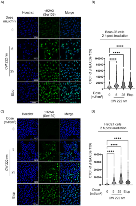
Cells had been seeded on coverslips and cultured for twenty-four h earlier than publicity to 222 nm mild, adopted by a further 2-h incubation. Cells had been then mounted in 4% paraformaldehyde for 15 min and incubated in a permeabilization/blocking buffer (1% BSA, 10% FBS, 0.1% Triton X-100, 0.1% Saponin) for 1 h at room temperature (RT).
Main antibody for γH2AX(Ser139) was diluted (1:200) and incubated in a single day at 4 °C, adopted by a 1-hour incubation with secondary antibody (Alexa FluorTM488 anti-Rabbit IgG, 1:1000) and Hoechst 33342 (1:1000) at RT in the dead of night. Samples had been then mounted and imaged utilizing a Leica SP8 confocal microscope with a 40× goal.
Fluorescence depth was quantified utilizing ImageJ software program, with corrected complete cell fluorescence (CTCF) calculated as: Built-in Density/(Cell Space × Imply Background Fluorescence).
Etoposide, a constructive management (a last focus of fifty µM, Etop), was used to induce DNA DSBs.
Immunofluorescence of formalin-fixed paraffin-embedded human trachea
Irradiated-human trachea samples and controls had been mounted in 4% impartial buffered formalin in a single day at RT, processed utilizing a regular automated tissue processor, and embedded in paraffin wax. Sections (5–8 μm) had been reduce for labelling experiments.
Slides had been dewaxed sequentially in xylene, ethanol gradients (100%, 90%, 70%, 40%), and dH2O, then transferred to citrate buffer (pH 6.0) for antigen retrieval through microwave heating (20 min). After cooling (30 min) and rinsing in working water (15 min), slides had been positioned in PBS to stop drying. Tissue sections had been framed with a PAP pen and blocked in a single day at 4 °C in PBS containing 1% BSA, 10% FBS, 0.1% Triton X-100, and 0.3% Saponin.
After washing with PBS, slides had been incubated with major antibody: anti-γH2AX(Ser139) or anti-(6 -4) DNA photoproducts in immunofluorescence buffer (PBS with 3% BSA, 1% goat serum, 0.1% Triton X-100, 0.1% Saponin) in a single day at 4 °C. Every major antibody was incubated with a unique tissue reduce. A small parafilm cowl was used to stop drying. After washing with 0.5% Tween 20 in PBS, slides had been incubated with secondary antibody (Alexa FluorTM488 anti-Rabbit IgG or Alexa FluorTM594 anti-Rabbit IgG, 1:1000, respectively) and Hoechst 33,342 (1:1000) for 1 h at RT in the dead of night.
Following washes with 0.5% Tween 20 in PBS, Vector TrueVIEW reagent was utilized (5 min) to scale back autofluorescence. Slides had been mounted with Vectashield Vibrance Antifade Mounting Medium and imaged utilizing a Leica SP8 confocal microscope with a 40x goal. Picture reconstruction was carried out with ImageJ software program.
Annexin V apoptosis detection
Irradiated cells and controls had been incubated in a humidified ambiance at 37 °C with 5% CO2 for 12 h. The medium was aspirated and preserved for the identification of apoptotic cells. The adherent cells had been washed with 2 mL of PBS. After washing, cells had been incubated with 0.5 mL of TrypLE Categorical/with out Phenol Pink for 1 min at RT. After including 2 mL of PBS, a cell suspension, together with non-adherent and adherent cells, was centrifugated at 400 g for five min. For every therapy 100 µL of cell suspension (1 × 10⁶ cells) was blended with FITC Annexin V (2.5 µL) and Propidium Iodide Resolution (1 µL, PI), a viability dye, and incubated for 15 min at RT shielded from mild. The cells had been washed with 400 µL of Annexin V Binding Buffer and centrifugated at 400 g for five min. The cells had been resuspended in 250 µL of Annexin V Binding Buffer and analyzed by Cytek Aurora (Cytek Biosciences). The stream cytometry information was analyzed by FlowJo software program. Staurosporine, a constructive management (a last focus of 1.25 µM, ST), was used to induce a non-lytic apoptosis.
Crystal violet assay
To evaluate the impact of Far-UVC publicity on cell viability, cells had been seeded into six-well plates at 0.35 × 10⁶ cells per effectively the day earlier than irradiation, reaching 70–80% confluence on the time of irradiation. Cells had been incubated in 500 µL of phenol red-free DMEM earlier than being uncovered to a particular Far-UVC dose and wavelength. After irradiation, the medium was changed with contemporary DMEM (2 mL per effectively), and cells had been incubated at 37 °C with 5% CO2 for the required period.
Cells had been then washed twice with PBS, mounted in 4% paraformaldehyde for 15 min at RT, and stained with 1 mL of 0.5% crystal violet (CV) in 20% methanol for 20 min at RT. After staining, cells had been washed 4 occasions with dH2O, air-dried, and solubilized in 1 mL of 30% acetic acid answer. The plate was incubated for 20 min with occasional shaking. Optical density (OD) was measured at 570 nm utilizing a Synergy H1 Hybrid Reader (BioTek Devices, Winooski, VT, USA). Etoposide (a last focus of fifty µM, Etop) was used as a constructive management.
Lactate dehydrogenase (LDH) cytotoxicity assay
Mobile LDH launched into the mobile medium upon plasma membrane harm by 222 nm mild was assessed with CyQuant LDH Cytotoxicity assay package in line with the producer’s directions. The absorbance of LDH (OD) was measured at 490 nm utilizing a Synergy H1 Hybrid Reader (BioTek Devices, Winooski, VT, USA). Etoposide (a last focus of fifty µM, Etop) was used as a constructive management.
Statistical evaluation
Statistical evaluation was carried out utilizing GraphPad Prism10. Statistical variations had been decided through one-way ANOVA with comparability primarily based on a management column. Unbiased experiments had been carried out a minimum of 3 times or in any other case as said. Outcomes are expressed because the imply ± SD. p-value p p p p